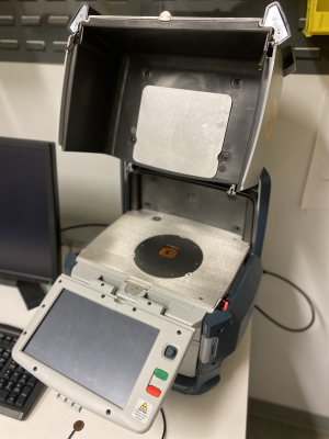
X-ray Fluorescence
| Niton FXL XRF [SOP] |
|---|
| Elemental Range: Mg and up (Z [math]\displaystyle{ \geq }[/math] 12) |
| *Mg – S (12 ≤ Z ≤ 16) unreliable in low concentrations |
| *Cl and up (Z ≥ 17) detectable in low concentrations |
| Chamber Dimensions: 9" (L) x 12" (W) x 4.5" (H) |
| Sample Type: Bulk, powders, & liquids/solutions |
X-ray fluorescence spectroscopy (XRF), also known as X-ray emission spectroscopy (XES), is a non-destructive characterization technique which gives elemental composition information. The MILL’s Niton FXL XRF accepts powdered, liquid, and bulk samples, and can detect elements heavier than magnesium ([math]\displaystyle{ Z\geq12 }[/math]). It is best suited for ceramic and metallic samples.
Operation
Anyone who wishes to use or be trained on the XRD must undergo the general lab safety training AND the Georgia Tech Office of Radiological Safety (ORS) X-Ray Safety Training before reporting to the XRD Technical Officer.
Theory
In an atom, electrons are bound to the nucleus with discrete binding energies—this is due to the quantized energy states we understand as “orbitals” or “shells.” When a material is struck by source radiation of sufficient energy, it dislodges a core electron, resulting in a vacancy. This vacancy now must be filled, or quenched, by a higher-energy electron. When quenched, the energy difference between the two electron levels is considered a surplus, and must be ejected either as a photon or electron of that specific energy difference. This specific energy difference is characteristic to each element, and the resulting signal is therefore indicative of a material’s elemental composition.
Fluorescence vs. Auger Behavior
The two aforementioned post-quench behaviors during are competitive and are the basis behind two different characterization techniques. If the surplus energy is ejected as a photon, it is called a secondary photon—this is also known as fluorescence. If ejected as an electron, it is called an Auger (oh-ZHAY) electron. The two corresponding techniques are subsequently called X-ray fluorescence spectroscopy (XRF) and Auger electron spectroscopy (AES), and both use X-rays as the source radiation. Note that XRF is the same thing as X-ray emission spectroscopy (XES), since the detector is measuring the count and intensity of secondary X-rays. Typically, fluorescence is favored in heavier elements, and Auger behavior is favored in lighter elements—this is part of the reason why our XRF (and all XRFs) struggle to detect lighter elements.
XRF vs. EDS
Note that XRF is a similar technique to the EDS/EDX (Energy Dispersive X-ray Spectroscopy) systems found in SEMs, but XRF is more bulk sensitive than EDS. EDS in SEMs generally use electron beam sources with small spot sizes (a couple nm in diameter), which then generates an interaction volume 1-2 microns in diameter where characteristic X-rays are emitted. XRF, however, will conduct the reading over a much larger "spot" area, with X-rays penetrating anywhere from several μm to a few mm in depth. This depth depends on the sample itself and the energy of the X-rays used.
Data
XRF data is presented as an intensity against energy (eV or keV) plot, where peak location (energy) determines the characteristic emission line represented, and relative peak heights (intensity) determines the a compositional ratio. Known electron binding energies and emission lines can be found in the X-ray data booklet, found here.
